
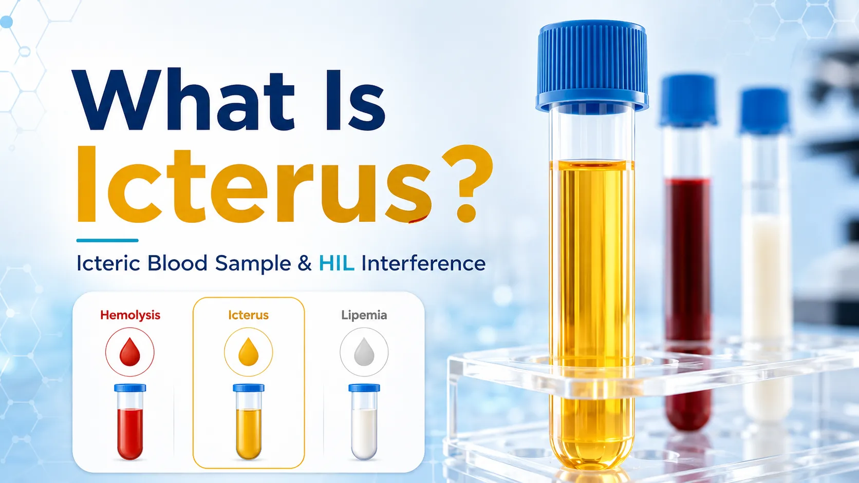
What is Icterus? Guide to Icteric Blood Sample & HIL Interference
Accurate laboratory results help doctors see what is happening inside your body so they can give you the right treatments. However, things can sometimes go wrong before your sample even reaches the laboratory machine.
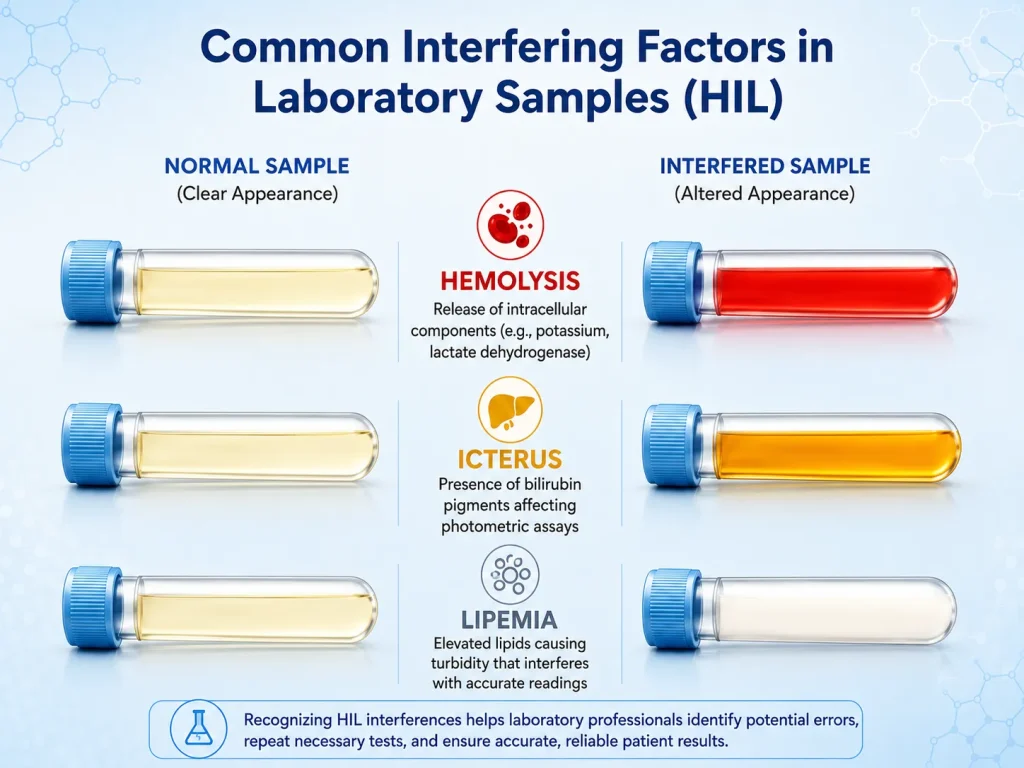
The three most common problems are Hemolysis, Icterus, and Lipemia. In the medical world, doctors call this HIL interference. When these issues happen, running an icteric blood sample or a hemolyzed specimen changes the color and clarity of your blood, causing lab machines to print out wrong or confusing numbers.
How HIL Interference Changes Lab Tests
Hemolysis: Breaks open red cells, spilling hidden chemicals into the liquid part of the blood.
Icterus: Adds a deep yellow bilirubin pigment that blocks the light paths inside testing machines.
Lipemia: Fills the blood with tiny fat droplets that scatter light and displace plasma water.
Understanding these three problems helps lab workers spot an icteric blood sample or a bad draw early so they can protect patient safety.
Understanding Hemolysis in Laboratory Samples
Hemolysis happens when red blood cells burst open. When they break, they spill a colorful protein called hemoglobin into the liquid part of the blood (the serum or plasma). This turns the normally clear, light-yellow sample into a pink or bright red color.
Hemolysis may happen inside the body (intravascular hemolysis) or outside the body (extravascular hemolysis) during blood collection or handling.
Common Causes of Hemolysis
Most of the time, cells burst by accident outside of the body while the blood is being collected or moved (in-vitro hemolysis):
- Using a needle that is too small or pulling the syringe plunger too hard.
- Shaking the blood tubes roughly instead of inverting them gently.
- Leaving blood tubes in places that are too hot or too cold.
- Waiting too long to separate the liquid blood from the cells.
⚠️In-Vivo Hemolysis: If cells burst inside the patient’s actual veins due to a disease (in-vivo hemolysis), a simple redraw will not fix the color. The lab must alert the doctor immediately to check for hemolytic anemia.
What is the Hemolysis Index?
The Hemolysis Index is a score given by automated machines to show exactly how many red cells broke open.
| Hemolysis Index Range | Interpretation |
|---|---|
| < 20 | No hemolysis (No broken cells) |
| 20–100 | Slightly hemolyzed specimen |
| 100–300 | Moderate hemolysis specimen |
| > 300 | Marked hemolysis specimen (Requires redraw) |
Lab Tests Affected by Hemolysis
Because red cells are full of minerals and proteins, bursting them open falsely changes your lab scores:
- Potassium: Falsely increased (Because potassium lives inside red cells).
- LDH & AST/ALT Enzymes: Falsely elevated.
- Bilirubin: Free hemoglobin blocks the machine’s light, causing a hemolyzed bilirubin testing clash.
⚠️ Important Note
Because of these distortions, laboratories usually request a new sample if hemolysis is significant.
How to Prevent Hemolysis When Drawing Blood
Best Practices:
- Use the correct needle gauge
- Gently invert tubes, avoid shaking
- Avoid drawing from IV sites
- Store samples promptly at recommended temperatures
- Separate plasma or serum quickly
Hemolysis Workup in Clinical Settings
A hemolysis workup helps determine whether the issue is in vivo (true hemolysis in the patient) or in vitro (sampling error).
- Intravascular hemolysis: Free hemoglobin in plasma, low haptoglobin, elevated indirect bilirubin
- Extravascular hemolysis: RBCs destroyed in the spleen/liver, increased bilirubin, normal plasma color
What is Icterus? Clinical Impact of an Icteric Blood Sample
By clinical definition, icterus means a dark yellow or brown discoloration caused by too much bilirubin pigment in the body. You might know this as jaundice, which turns the skin and the whites of a person’s eyes yellow. In the laboratory, processing an icteric blood sample means dealing with icteric serum or icteric plasma, which looks like dark amber juice instead of pale yellow can interfere with photometric tests because bilirubin absorbs light in similar wavelengths as many chemical reagents.
Clinical Causes of an Icteric Blood Sample
When a lab tech processes an icteric blood sample meaning high bilirubin levels are present, it usually points to one of these conditions:
- Liver damage or diseases like hepatitis or cirrhosis.
- Gallbladder or bile duct blockages that stop bile flow.
- Neonatal jaundice (When newborn babies have a temporary, immature liver).
What is the Icteric Index?
The Icteric Index indicates the concentration of bilirubin levels in the liquid portion of a blood sample.
| Icteric Index Range | Interpretation |
|---|---|
| < 1.0 mg/dL | Non-icteric (Normal) |
| 1.0 – 2.0 mg/dL | Mild icterus (Slight interference) |
| > 3.0 mg/dL | Severe icterus (Unreliable results) |
How Does an Icteric Sample Affect Lab Results?
An icteric blood sample causes severe errors because bilirubin absorbs light at the exact same wavelengths (450 nm) that lab machines use to read chemistry assays. This optical block can falsely lower important test numbers like glucose (sugar), cholesterol, and creatinine.
Managing Icterus Interference
Labs can reduce interference by:
- Using dual-wavelength (bichromatic) analysis
- Diluting samples to reduce bilirubin concentration
- Applying software corrections in automated systems
Icterus in Newborns (Neonatal Jaundice)
Neonatal jaundice is a frequent cause of icterus in newborns. The infant’s immature liver struggles to process bilirubin efficiently, leading to yellowing and possible lab result interference. Phototherapy helps break down bilirubin, and monitoring is crucial to ensure that test results reflect the baby’s true health status.
Understanding Lipemia in Blood Tests
Lipemia means there is too much fat in the blood. It makes the serum or plasma look cloudy, hazy, or milky-white instead of clear.
It primarily occurs after a fatty meal or in individuals with hyperlipidemia. Lipemia affects spectrophotometric tests by scattering light and diluting the plasma water phase.
Clinical Causes of Lipemia
- Non-fasting samples: Eating a heavy, fatty meal right before a blood draw.
- Hypertriglyceridemia: A medical condition where a person’s body makes too many lipids (fats).
- IV lipid infusions: Getting fat-based nutrition through an intravenous tube in the hospital.
What is the Lipemia Index?
Labs use the Lipemia Index to measure sample cloudiness (turbidity).
| Lipemia Index Range | Interpretation |
|---|---|
| < 60 | Clear sample (non-lipemic) |
| 60 – 120 | Slightly turbid (mild lipemia) |
| > 120 | Cloudy or milky sample (marked lipemia) |
How Lipemia Interferes with Laboratory Assays
Lipemia creates a problem called light scattering. The fat droplets act like tiny mirrors, bouncing the machine’s light rays in all directions so the sensors cannot read the absorbance properly.
Fat also takes up too much physical space in the tube. This causes a mistake called the volume displacement effect. The large lipid particles crowd out the liquid phase, forcing the machine to falsely report low numbers for electrolytes like sodium (Na+) and chloride (Cl-).
Methods for Reducing Lipemia Interference
How do labs fix this milky, fatty blood problem? They use three simple tricks to get clean and correct test results:
- Patient Fasting: The easiest way is to ask the patient not to eat anything for 10 to 12 hours before their blood test. When you do not eat, your body naturally clears away the fat from your last meal, making the blood clear again.
- Ultracentrifugation (Super Spinning): Imagine putting the cloudy blood sample into a super-fast spinning machine. This machine spins so hard that the heavy fat droplets float straight to the top. After that, the lab worker can easily skim the fat off, leaving clean blood at the bottom for testing.
- Serum Blanking (Color Subtracting): This is like a smart computer trick used in spectrophotometric tests (tests that measure light). The laboratory machine shines a light and takes a reading of the cloudy, fatty blood first to measure the “background noise.” Then, it subtracts that cloudiness from the final score, allowing the machine to see the real, true test color.
How to Spot HIL Interference at the Bench
Lab workers must examine samples visually after spinning them down in a centrifuge. Recognizing these physical changes immediately tells the technician if a test needs special handling.
When looking at blood tubes on the work bench, a normal sample looks clear and light yellow. In contrast, a hemolyzed sample turns a clear pink or deep red, a lipemic specimen looks thick and milky-white, and an icteric blood sample turns a very dark amber-yellow.
Frequently Asked Questions (FAQ)
Q1: What is hemolysis, icterus, and lipemia?
Hemolysis means broken red blood cells turn the sample red. Icterus means high bilirubin turns the sample dark yellow. Lipemia means high fat turns the sample milky-white. All three cause HIL lab errors.
Q2: What causes hemolysis interference in laboratory samples?
Ans: Hemolysis interference is usually caused by accidental damage to red blood cells during or after blood collection (in-vitro hemolysis). The most common causes include:
- Incorrect Needle Size: Using a needle that is too small, which forces blood cells through a tight space and causes them to burst.
- Rough Handling: Shaking or mixing the blood collection tubes too aggressively instead of inverting them gently.
- Temperature Extremes: Leaving the blood samples in places that are too hot or too cold.
- Processing Delays: Waiting too long to separate the liquid serum or plasma from the actual blood cells.
Q3: What causes lipemia in a blood test?
The most common cause is eating fatty foods right before your blood draws. It can also be caused by genetic conditions, diabetes, or medication side effects.
Q4. What does it mean when a sample is icteric?
An icteric blood sample is caused by a buildup of bilirubin. This happens when the liver cannot clear broken-down red blood cells properly, or when the bile ducts are blocked.
Q5: What is lipemic blood?
Lipemic blood is blood that looks cloudy or milky because it is crowded with large fat particles. A simple 12-hour fast before your blood test usually prevents this problem.
Conclusion
In laboratory medicine, accurate numbers save lives. Hemolysis, icterus, and lipemia remain the biggest daily challenges to getting those numbers right. By tracking HIL indices, utilizing proper blood draw steps, and running sample-clearing techniques, lab professionals can stop pre-analytical errors when processing an icteric blood sample or turbid specimen.
For more insights on clinical laboratory best practices and pre-analytical quality control, MFZ Clinical Lab provides comprehensive educational resources for laboratory professionals.
References:
Joe M. El-Khoury. Hemolysis, Icterus, and Lipemia Interference: New Approaches to Old Foes. [Clinical Lab]
Alain Nicolay, Anne‐Marie Lorec, Guy Gomez, Henri Portugal. Icteric human samples: Icterus index and method of estimating an interference‐free value for 16 biochemical analyses. [PMC free article]
Medical Disclaimer: The information provided on MFZ Clinical Lab is for educational and informational purposes only. It is intended to help students and laboratory professionals understand clinical concepts and should not be used as medical advice, diagnosis, or treatment. Always consult a qualified healthcare provider or doctor for any medical concerns or interpretation of lab test results.
